Teleradiology
Reimagined.
Empower your remote reading practice with the fastest viewer, seamless dictation, and intelligent worklist management – all in one platform.
Omni-Box
Dictate freely & AI distributes to sections – or chat with AI.
Radiologist-First Platform
Designed by radiologists & developers together, our platform is optimized for a radiologist's every need.
2x Faster Reports
AI-powered dictation eliminates navigation commands & automates impressions so you can focus on diagnosis.
Multiple PACS, One Worklist
Add new imaging center partners in minutes. See all studies in one unified worklist.
Per-Study Pricing
No seat licenses. Costs scale with volume. Transparent billing you can track by partner.
Read our blog on why AutoScribe is a great fit for radiology groups.
Read hereEverything You Need to
Read Faster.
AutoScribe's feature suite is designed to eliminate friction at every step of the reporting workflow.
liver normal size, spleen unremarkable, kidneys show cortical thinning bilaterally...
Omni-mode merges:
→Liver: Normal in size, no focal lesions...
→Spleen: Unremarkable...
→Kidneys: Cortical thinning bilaterally...
→Gallbladder: Surgically absent...
→Pancreas: Unremarkable...
Recommend repeat CT chest in 3 months to assess nodule stability.
MRI liver with contrast advised for further characterization.
Moderate 60% stenosis in mid-RCA with calcified plaque.
Mild 35% stenosis in proximal LAD with mixed plaque composition.
Pericoronary adipose tissue attenuation within normal limits.
AI Impressions, Follow-up, & Comparison
Single click impressions, follow-up, and comparison direct from your findings.
2.1 cm hypoechoic nodule in the right thyroid lobe with irregular margins and microcalcifications. TI-RADS 4, recommend FNA biopsy. Left thyroid lobe unremarkable.
Classifications
Severity Classifications
Color-coded findings let referring providers triage critical results instantly.
Components
Divider
Drag to add
Section
Drag to add
Description
Drag to add
Macro
Drag to add
Template Preview
• Normal size and echotexture
• No focal lesions identified
• No cholelithiasis
• Wall thickness normal
• Bilateral normal size
• No hydronephrosis
• No suspicious masses
• Normal fibroglandular density
• Normal size
• Homogeneous echotexture
Explore more on how AutoScribe can accelerate your workflow.
Learn moreMultiple PACS,
One Unified Worklist
Connect to multiple imaging center partners with dedicated API keys. All studies flow into a single, aggregated worklist – no tab switching, no missed reads.
Schedule DemoWorklist
Unified View
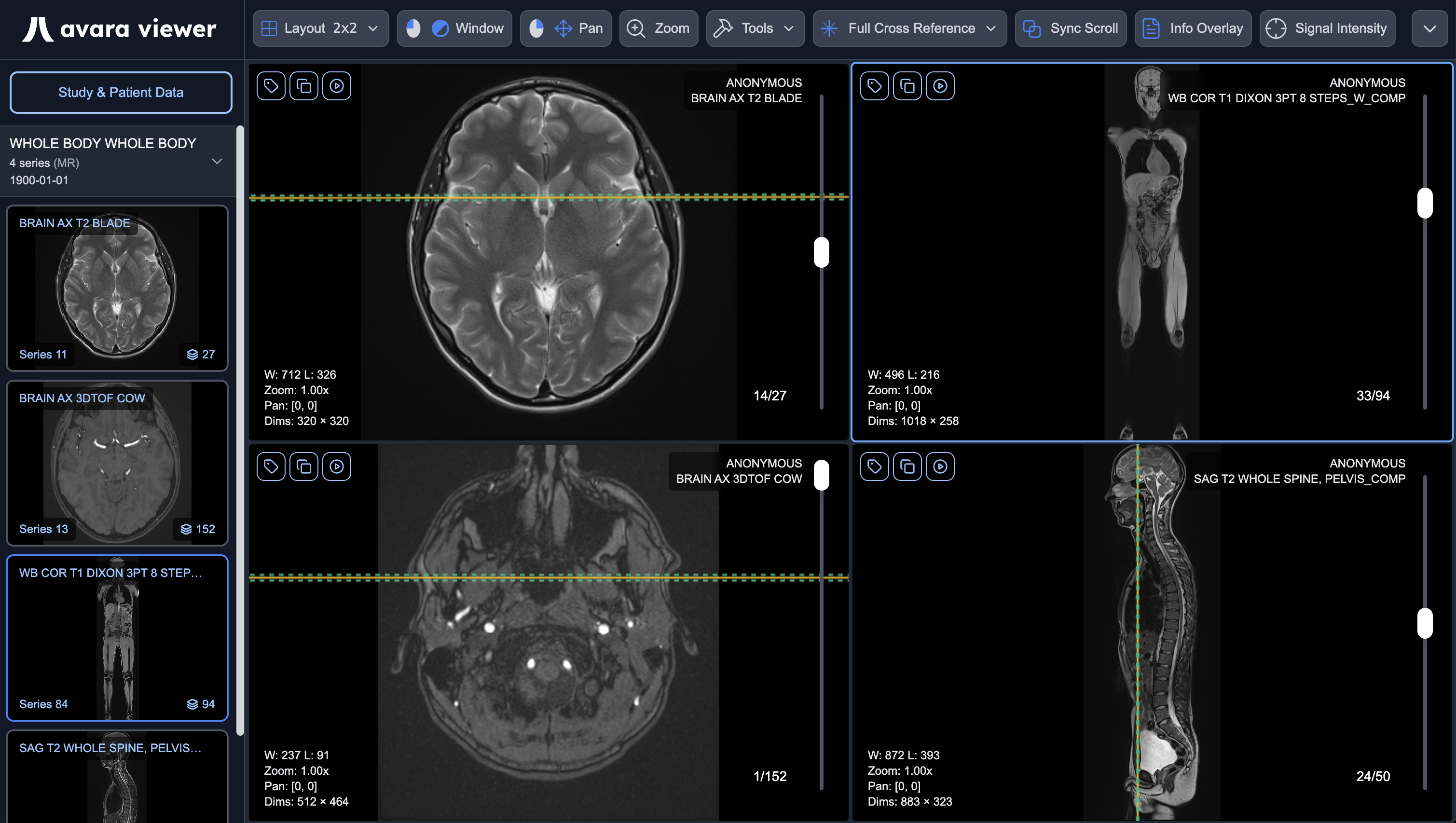
A Diagnostic Viewer
Included at No Cost
Avara's web-based DICOM viewer is built into every study as an optional enhancement. Read studies directly in-platform – no additional software licenses required.
Accelerate Your Teleradiology Workflow
Join teleradiology groups across the country delivering faster, more accurate reports.
Frequently Asked Questions
Yes. AutoScribe is designed to integrate seamlessly with PACS, RIS, and EHR systems through our SDK. PACS and EHR companies can also offer AutoScribe as a Software-as-a-Service product to their customers using our highly programmable SDK.
AutoScribe uses per-study pricing – you only pay for what you read. There are no seat licenses, no monthly minimums, and no long-term contracts. This model is ideal for teleradiology groups with variable volume across multiple partners.
Yes. Avara Viewer is included free when you use AutoScribe or our Clinical Platform.
AutoScribe achieves 96.6% accuracy – the highest in the industry with the lowest recorded error rate on the market. Our AI continuously learns and improves based on your dictation patterns.
Yes. We offer a 30-day free trial so you can experience AutoScribe's features and see how it transforms your workflow before committing.