The Fastest Diagnostic
ViewerEver Built.
FDA-cleared, GPU-optimized, web-based DICOM viewing software to deliver the best possible reading experience.
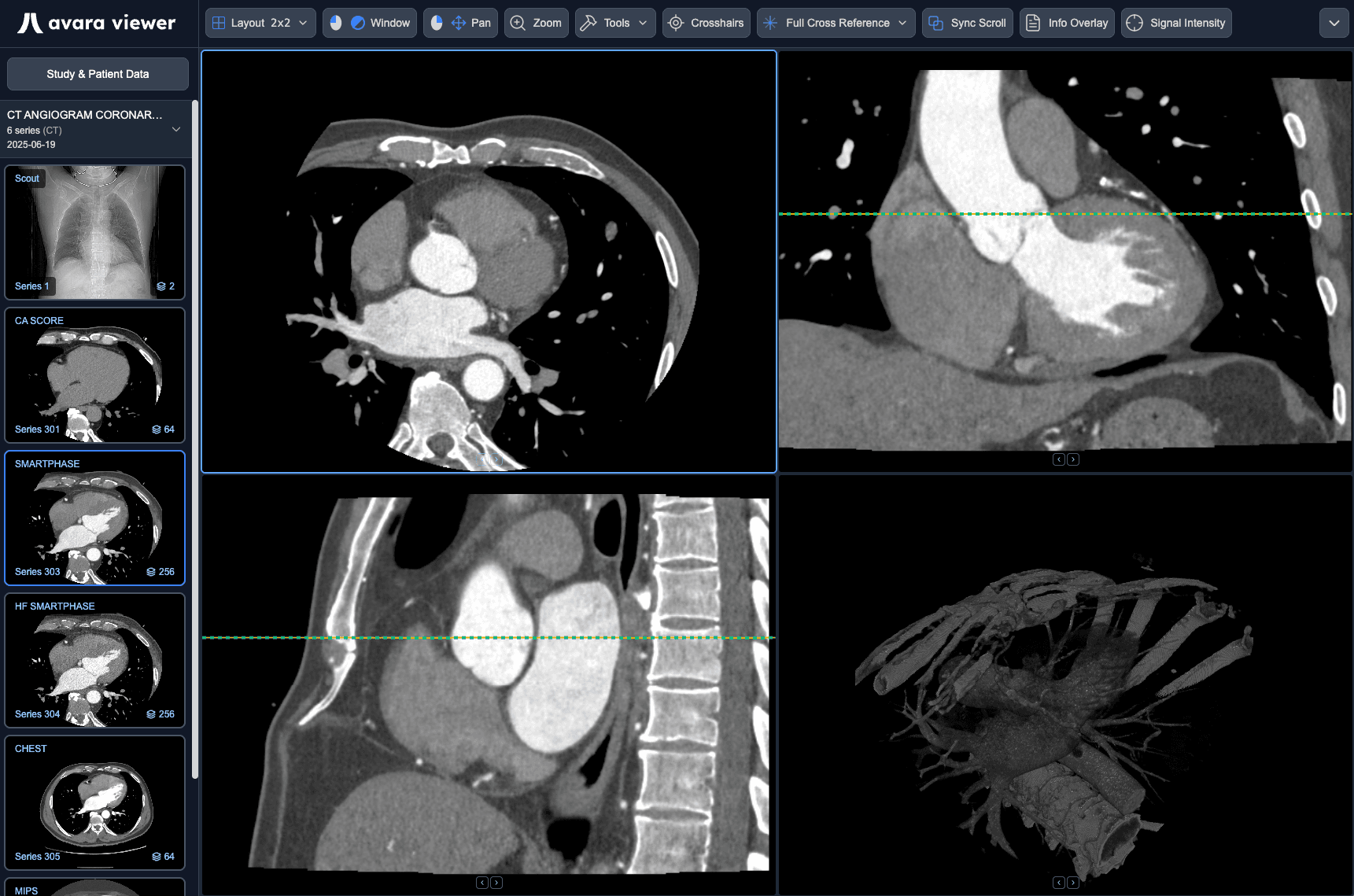
Zero-lag reading
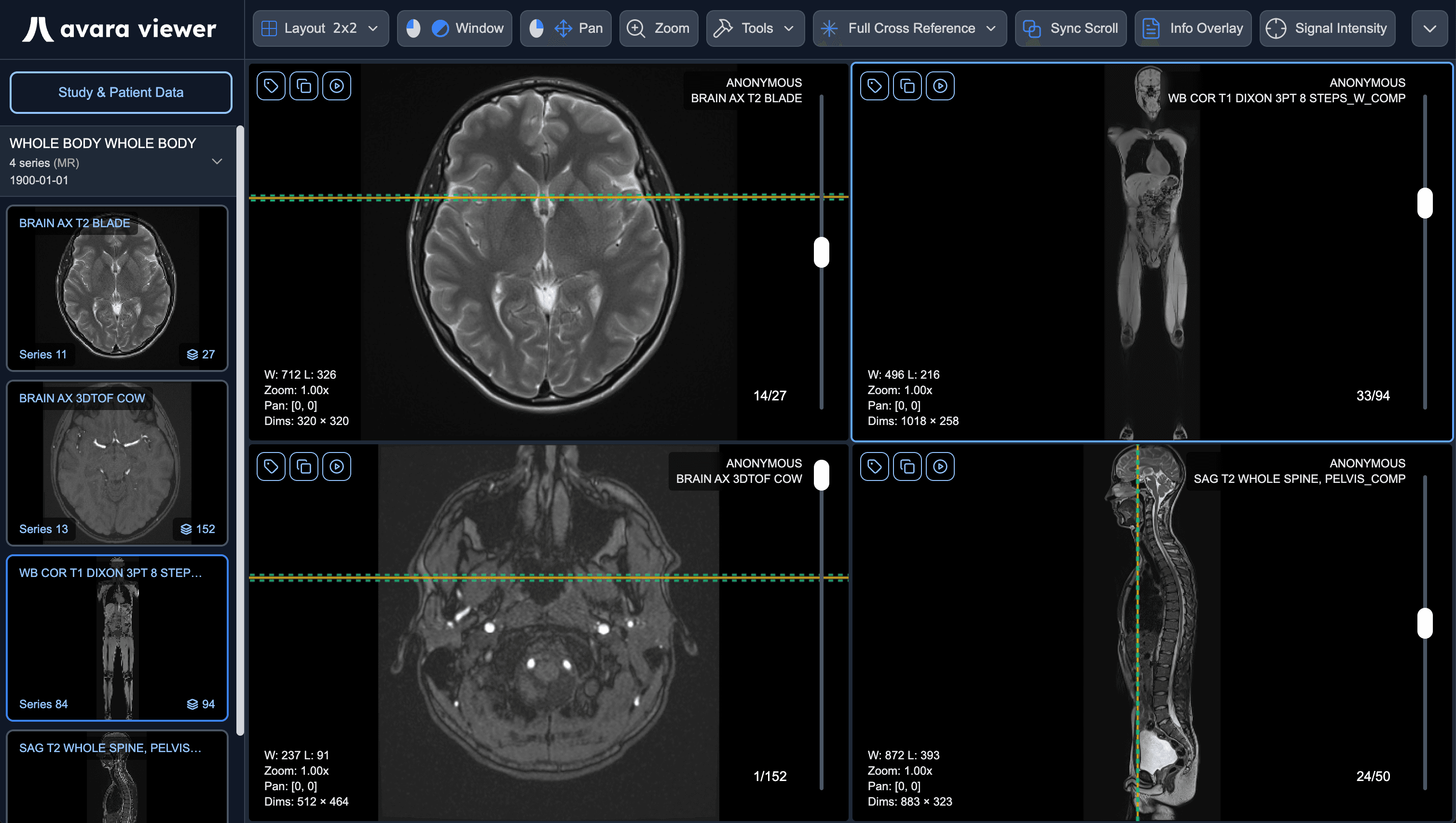
Supports all modalities
Fast. Elegant. Intuitive.
Ultra-optimized web-based DICOM viewer that lets you
read with clarity & precision.
3D Rendering in
Under a Second
Transform any DICOM modality into an interactive 3D model with a single click.
Imaged to the right is a Circle-of-Willis MRI time-of-flight sequence rendered in the Avara Viewer.
Fluid Interaction.
No Lag – Ever.
Our viewer is GPU-optimized at every turn to deliver cross-reference & viewport scroll syncing with absolutely zero lag.
Schedule DemoComprehensive Tool Suite to Meet Every Reader's Needs
Everything you need for efficient, accurate diagnostic reading – built directly into your workflow.
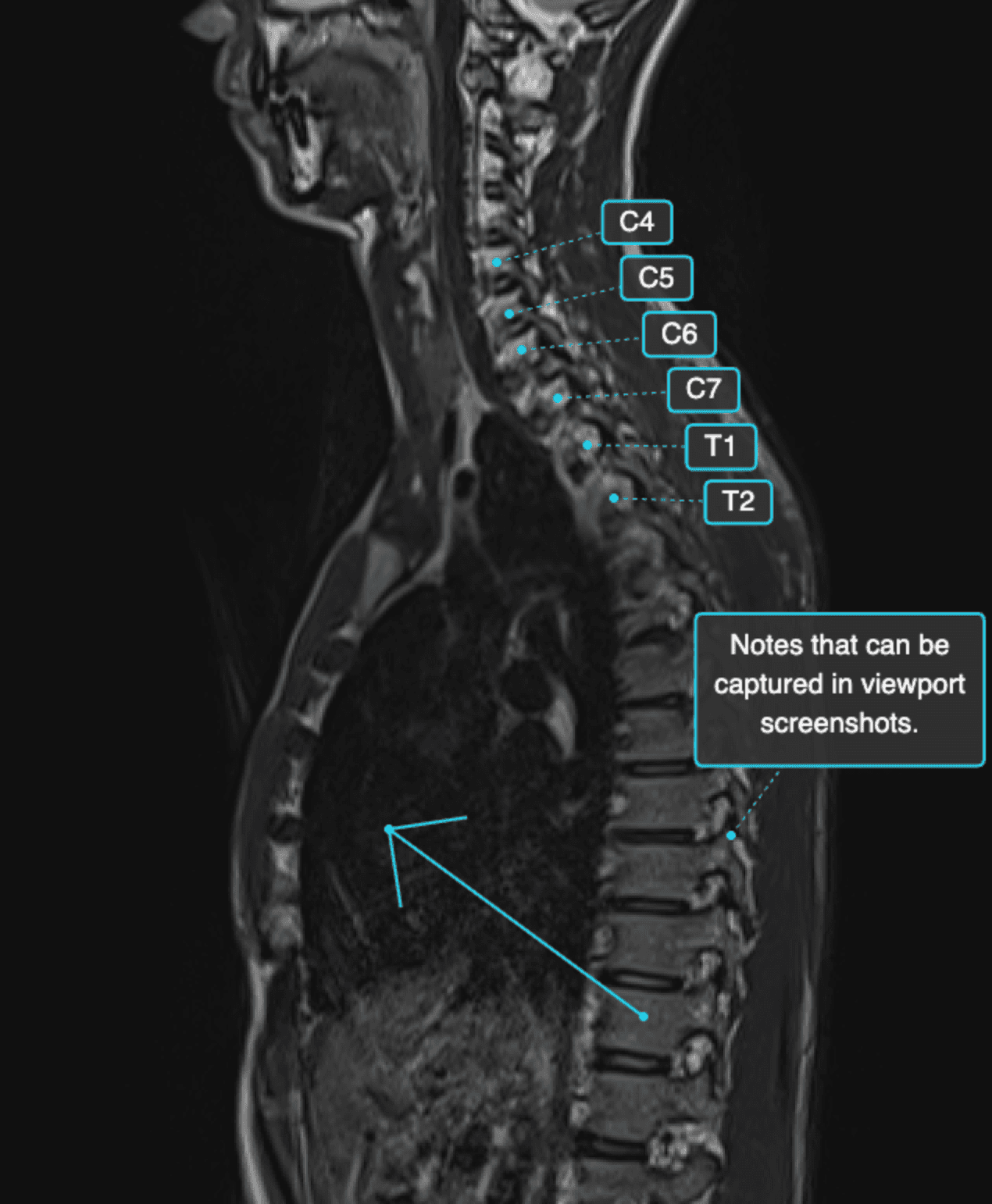
Annotate with Confidence
Enhance reading & viewport annotations with vertebra labels, text-boxes, arrows, and pencil.
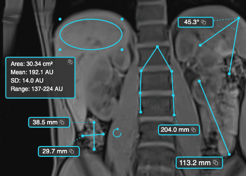
Measure with Precision
Provide diagnostic measurements with our line, angle, ROI, lesion bidirectional, polyline, and recalibration tools.
Visualize Blood Flow
Play through sequences like a movie with the cine tool – frame rates up to 40 FPS supported.
MIP Vascular Studies
With the click of a button, toggle maximum intensity projection and adjust the width with ease.
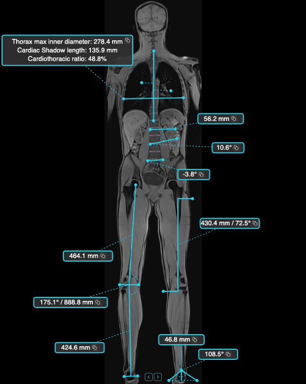
Specialty Measurement Tools
Advanced specialty tools for musculoskeletal and related workflows – height difference, goniometry, foot longitudinal arch, cardiothoracic ratio, Cobb angle, and vertebra angle.
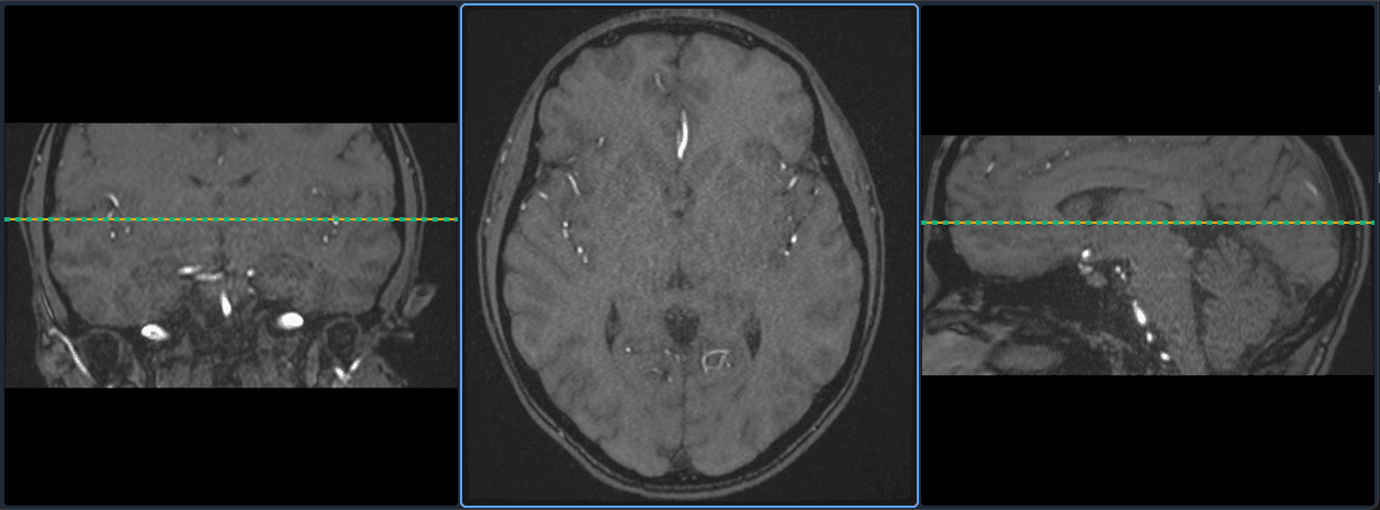
Project Into Any Plane
Advanced multiplanar reconstruction rendering in under a second – view any series in axial, coronal, or sagittal planes instantly.
Full Mammography &
Tomosynthesis Support
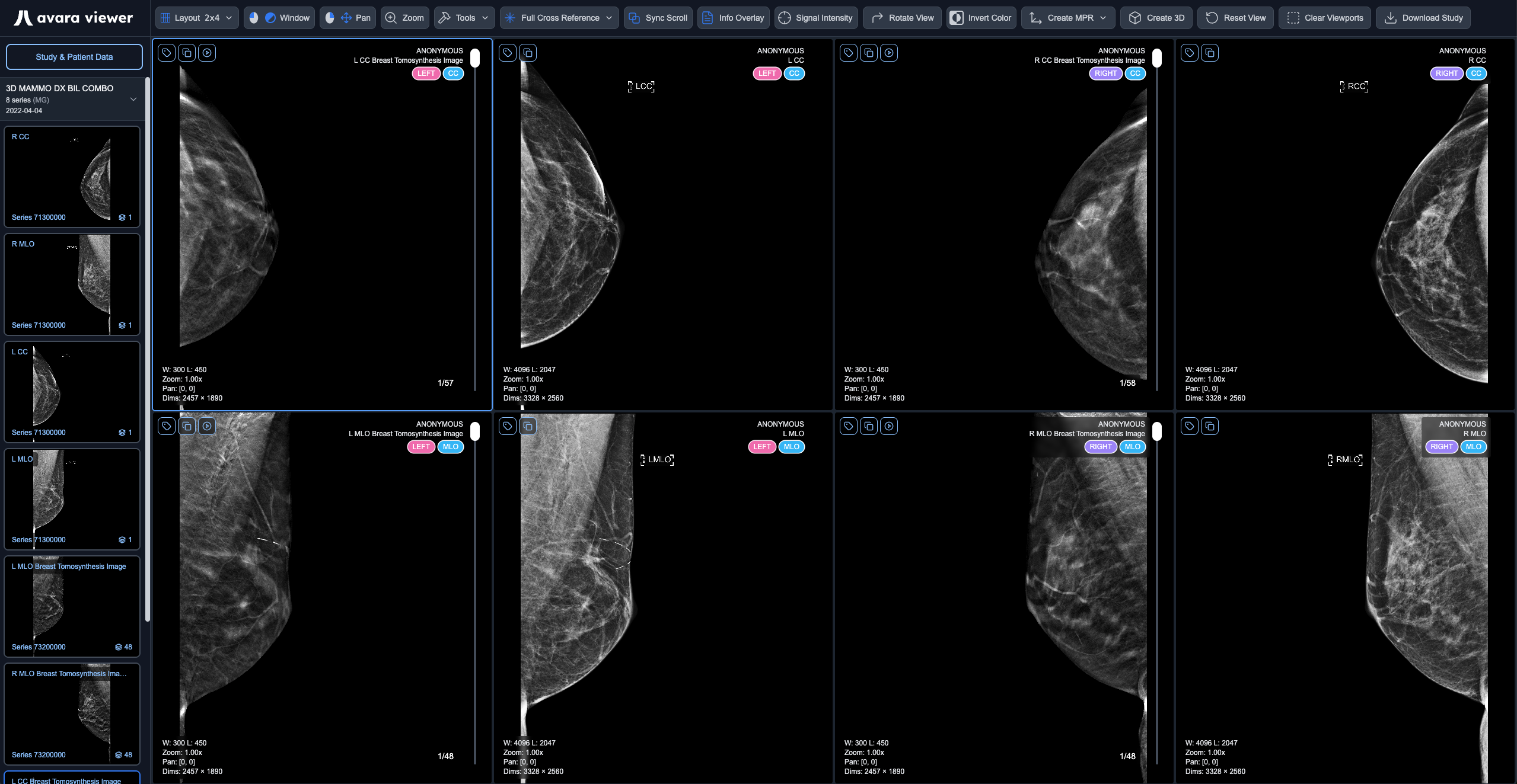
Mammo & DBT studies automatically render a predictable hanging protocol on load that follows an intuitive & powerful rules engine.
Learn More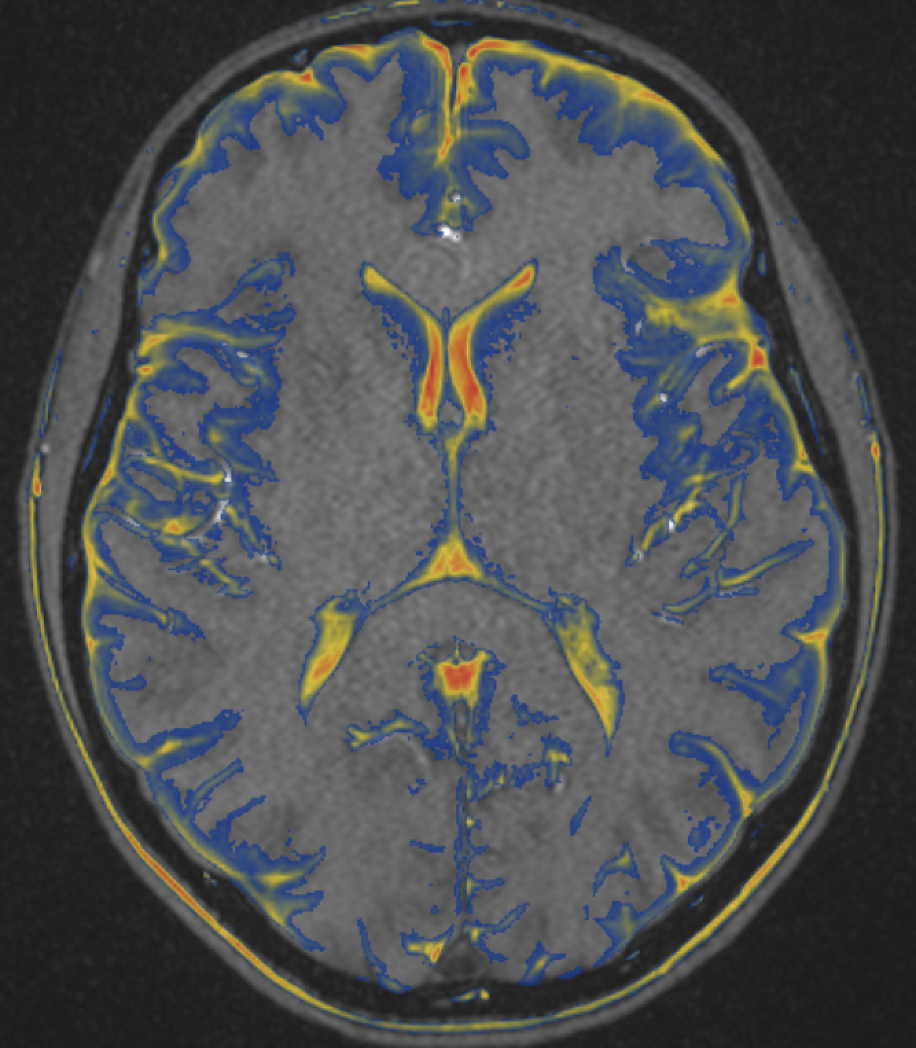
Fusion for
Any Modality
Seamlessly overlay PET, SPECT, or any functional imaging data onto CT or MRI anatomical studies for enhanced diagnostic precision.
QT Ultrasound Module
QT Ultrasound studies automatically render a dedicated hanging protocol with quantitative FGV, TBV, and FGR density overlays.
Learn More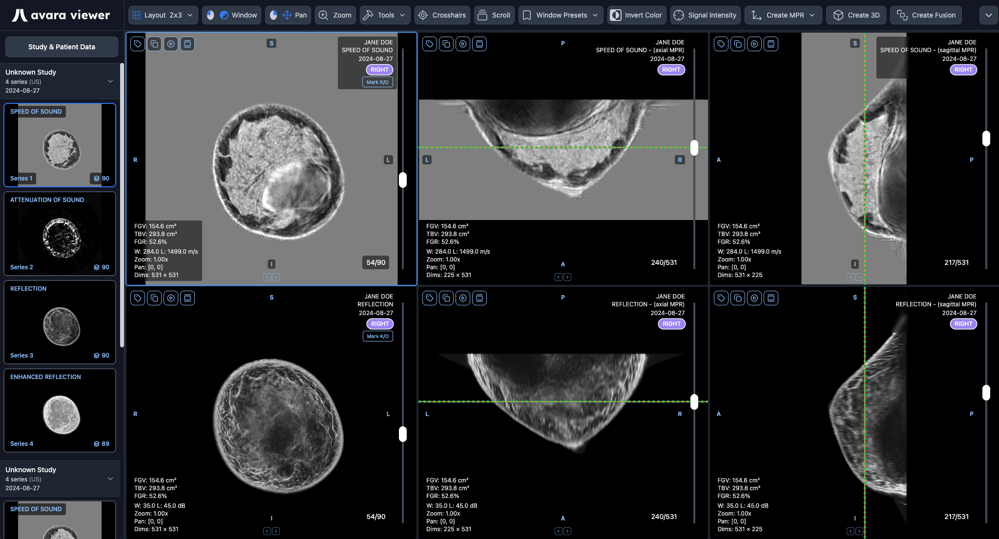
Connect any PACS to the Avara Viewer. Add and remove sites seamlessly, with no interfacing or setup fees.
Avara PACS
connected
Northline PACS
connected
Viewer Worklist
Live18 studies ready to read
Leah Kim
CT Angiogram · Northline
Theo Marquez
MRI Lumbar · Avara PACS
Isla Bennett
CT Abdomen · Fairmont
Owen Drake
XR Knee · Cedar Ridge
Maya Singh
US Thyroid · Redwood
Fairmont RIS
connected
Redwood Imaging
connected
Transform How You Read
Trusted by practices across the country to deliver faster,
more accurate reads.
Frequently Asked Questions
Yes. Avara Viewer has received FDA clearance under K number K262120 as a Class II medical device for diagnostic use across all modalities, including mammography. There are no modality restrictions – it's fully cleared for diagnostic interpretation.
Yes. Powerful hanging protocols can be configured for different study types and appointment types, spanning multi-monitor and multi-viewport setups – including toolbar configuration for each hanging protocol. Automatic mammography hanging protocols choose layouts based on laterality, view, and display tags. Our team is happy to help you set up protocols for your readers.
Yes. Common viewport and toolbar actions have keyboard shortcuts, including:
- Up/Down arrows: navigate frames in the active series
- Left/Right arrows: move to the previous or next series in the active viewport
- Delete / Backspace: delete the selected annotation
- Esc: cancel an in-progress annotation
Toolbar actions are also split into continuous tools (window, pan, zoom, measure, annotate) and discrete actions (layout, MPR, 3D, fusion, invert, reset, download) so bindings stay predictable. Tools and settings can also be configured with customizable keyboard shortcuts so each reader can bind the actions they use most.
8GB of RAM is the absolute minimum for diagnostic use. Below 8GB you should expect severe performance degradation.
For production reading, plan from how you actually work: number of simultaneous viewer instances × largest anticipated study size (GB) × 6. Example: 2 instances and a 500MB study → 2 × 0.5 × 6 = 6GB of workload headroom, so the recommended system target remains the 8GB floor. Memory-intensive workflows (large CT/MR, tomosynthesis, multiple monitors) may need 16GB, 32GB, or more.
Yes. Avara Viewer is included free when you use AutoScribe or our Clinical Platform.